Help yourself to learn the anatomy of the teeth structure using these free and printable labeled diagrams! These 101 Diagramss are designed to guide you in studying the structure of human teeth. We use our teeth to break down items of food by cutting and crushing them in preparation for swallowing and digesting. Humans have four types of teeth: incisors, canines, premolars, and molars, each with a specific function. Take a look at the following teeth diagram of adult provided below.

In the diagram of teeth above, you can see the illustration of adult teeth. The type, number, and arrangement of a set of teeth represent the dentition. In the teeth structures, the incisors cut the food, the canines tear the food and the molars and premolars crush the food. The roots of teeth are embedded in the maxilla (upper jaw) or the mandible (lower jaw) and are covered by gums. The tooth has two anatomical parts. The crown of a tooth is that part of the tooth which is covered with enamel and this is the part usually visible in the mouth. The root is the part embedded in the jaw. It anchors the tooth in its bony socket and is normally not visible. More structure diagrams of the teeth are posted below.


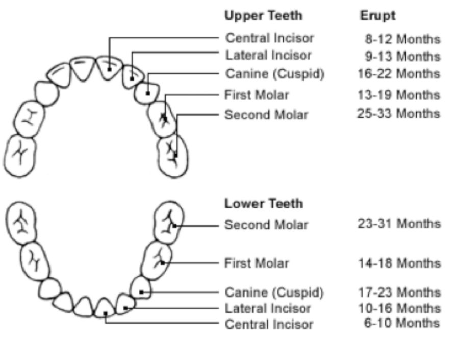
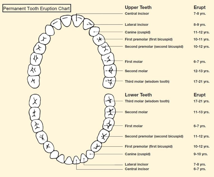
A normal adult mouthhas 32 teeth, which (except for wisdom teeth) have erupted by about age 13. The crown of each tooth projects into the mouth. The root of each tooth descends below the gum line, into the jaw. Humans have two set of teeth, primary and permanent teeth. Primary teeth start to form during the embryo phase and erupt during infancy (from 6 months to 3 years). On the other hand, the first permanent teeth appear around the age of 6.

All these pictures presented are printable teeth diagram resources. Learn more about the other educational diagrams by browsing through our categories or looking it up on the search column!